
Cervical Lymph Node Levels
|
Level I |
Submental and submandibular nodes. |
| Level IA | Submental nodes, between the medial margins of the anterior bellies of the digastric muscles. |
| Level IB | Submandibular nodes, lateral to level IA nodes and anterior to the back of the submandibular salivary gland. |
| Level II | Upper internal jugular nodes, posterior to the back of the submandibular salivary gland, anterior to the back of the sternocleidomastoid muscle and above the level of the bottom of the body of the hyoid bone. |
| Level IIA | Nodes lie posterior to the internal jugular vein and are inseparable from the vein, or they are nodes that lie anterior, medial, or lateral to the vein. |
| Level IIB | Nodes lie posterior to the internal jugular vein and have a fat plane separating the nodes and the vein. |
| Level III | Middle jugular nodes, between the level of the bottom of the body of the hyoid bone and the level of the bottom of the cricoid arch, anterior to the back of the sternocleidomastoid muscle. |
| Level IV | Low jugular nodes, between the level of the bottom of the cricoid arch and the level of the clavicle, anterior to a line connecting the back of the sternocleidomastoid muscle and the posterolateral margin of the anterior scalene muscles; they are lateral to the carotid arteries. |
| Level V | Posterior triangle nodes, posterior to the back of the sternocleidomastoid muscle, and posterior to the line described in level IV. |
| Level VA | Above the level of the bottom of the cricoid arch. |
| Level VB | Between the level of the bottom of the cricoid arch and the level of the clavicle. |
| Level VI | Upper visceral nodes, between the carotid arteries from the level of the bottom of the body of the hyoid bone to the level of the top of the manubrium. |
| Level VII | Superior mediastinal nodes, between the carotid arteries below the level of the top of the manubrium and above the innominate vein. |
| Supraclavicular nodes | Nodes at, or caudal to, the level of the clavicle and lateral to the carotid artery. |
| Retropharyngeal nodes | Nodes behind the pharynx, medial to the internal carotid artery, from the skull base down to the level of the hyoid bone. |

Figure 1. Diagram of the neck as seen from the left anterior view. Left, The pertinent anatomy that relates to the nodal classification. Right, An outline of the levels of the classification. Note that the line of separation between levels I and II is the posterior margin of the submandibular gland. The separation between levels II and III and level V is the posterior edge of the sternocleidomastoid muscle. However, the line of separation between levels IV and V is an oblique line extending from the posterior edge of the sternocleidomastoid muscle to the posterior edge of the anterior scalene muscle. The posterior edge of the internal jugular vein separates level IIA and IIB nodes. The top of the manubrium separates levels VI and VII.
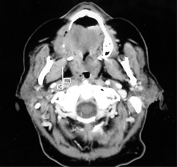
Figure 2. Axial postcontrast computed tomographic scan of the neck through the level of C2 shows a sagittally oriented white line drawn along the medial edge of the internal carotid artery (IC). Within 2 cm of the skull base, any node medial to this line is classified as a retropharyngeal node, while a node lateral to this line is a level II node. RN indicates a necrotic lateral retropharyngeal node.
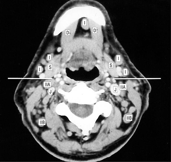
Figure 3. Axial postcontrast computed tomographic scan of the neck just cranial to the hyoid bone. A transverse white line has been drawn through the posterior edge of each submandibular gland (S). The nodes anterior to this line are level I nodes, while the nodes posterior to this line are level II nodes. The node between the anterior bellies of the digastric muscles (D) can be subclassified as level IA, while the other level I nodes lateral to the medial edge of each anterior digastric belly can be subclassified as level IB nodes. The level II nodes anterior to and/or touching the internal jugular vein (J) are subclassified as level IIA. Those level II nodes posterior to the vein and not touching it are subclassified as level IIB.
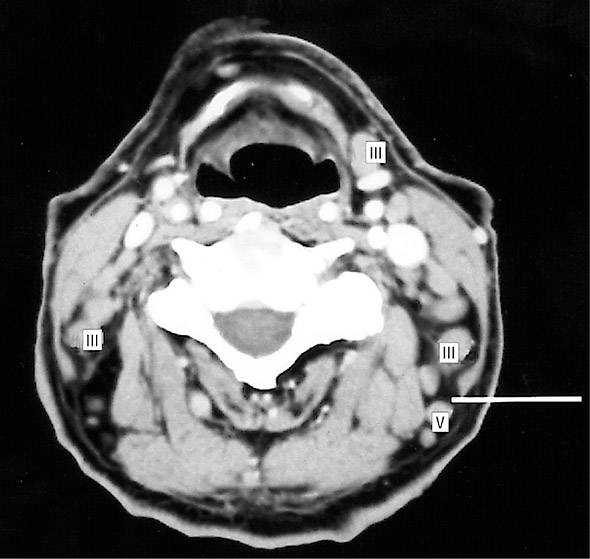
Figure 4. Axial postcontrast computed tomographic scan of the neck just caudal to the hyoid bone. A transverse white line has been drawn through the posterior edge of the left sternocleidomastoid muscle. Note that level III nodes lie anterior to this line and include those nodes anterior to the carotid sheath structures (internal jugular nodes) as well as those deep to the sternocleidomastoid muscle (spinal accessory nodes). Posterior to the transverse line are level V nodes.
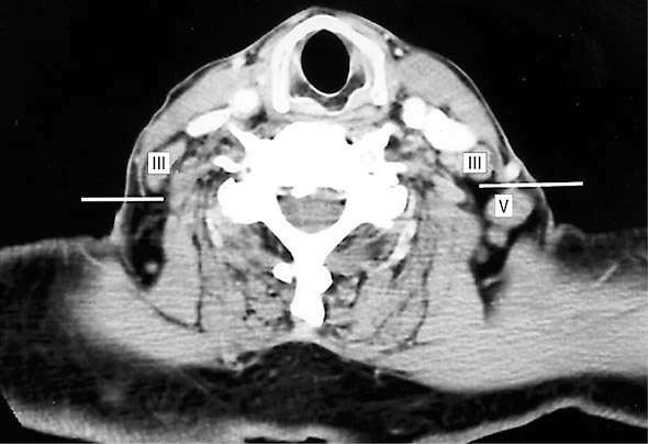
Figure 5. Axial postcontrast computed tomographic scan of the neck through the level of the cricoid cartilage, cranial to the lower border of the anterior cricoid arch. A transverse white line has been drawn through the posterior edge of each sternocleidomastoid muscle. Nodes anterior to this line are level III, while nodes posterior to this line are level V.
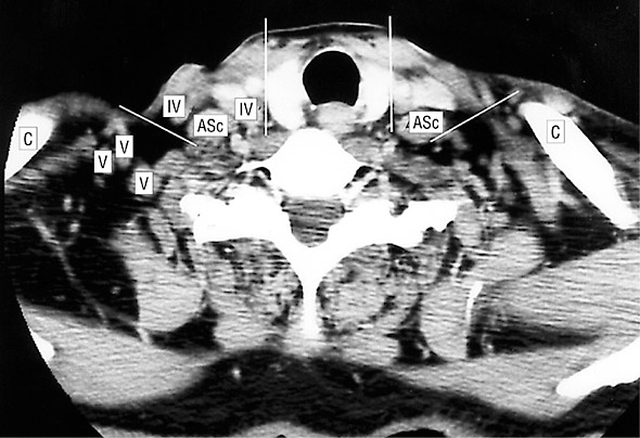
Figure 6. Axial postcontrast computed tomographic scan of the neck through the thyroid gland, caudal to the lower border of the cricoid cartilage arch. Oblique white lines on each side have been drawn from the posterior edge of the sternocleidomastoid muscle to the lateral posterior edge of the anterior scalene muscle (ASc). Vertical lines have also been drawn from the medial edge of each common carotid artery. Nodes anterior to the oblique lines and lateral to the vertical lines are level IV nodes. The nodes posterior to the oblique lines are level V nodes. Level VI nodes are between the common carotid arteries (vertical lines). Note that the level IV nodes in this figure are the prescalene nodes described by Rouviere2 and others.3-4 The clavicle (C) is identified for reference. The level V nodes in this figure should actually be classified as supraclavicular nodes, as they are at and caudal to the visualized portion of the clavicle.
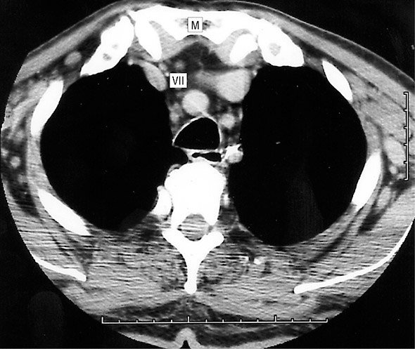
Figure 7. Axial postcontrast computed tomographic scan through the level of the top of the manubrium (M). Caudal to the top of the manubrium, the visceral nodes are classified as level VII. Cranial to this level, the visceral nodes are level VI nodes.
Reference:
An Imaging-Based Classification for the Cervical Nodes Designed as an Adjunct to Recent Clinically Based Nodal Classifications Peter M. Som, MD; Hugh D. Curtin, MD; Anthony A. Mancuso, MD, Arch Otolaryngol Head Neck Surg. 1999;125:388-396. PDF