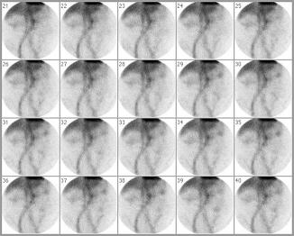
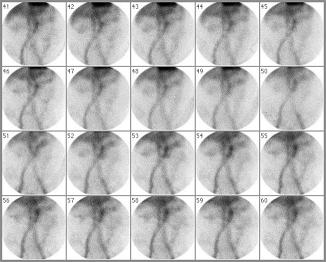
The patient is a 79-year-old male with hematochezia who has had a negative upper GI and lower GI endoscopy.


After 15 min a blush of tracer activity is seen in the transverse colon, just next to the midline, near the region of the splenic flexure. This diffuse area of abnormal activity migrates to the left splenic flexure and then inferiorly towards the left lower quadrant along the expected anatomy of the descending colon. Normal tracer activity is seen in the blood pool of the abdominal aorta, common iliac arteries, femoral arteries, liver and spleen on all of the interval images.
A focus of abnormal RBC pooling is seen in the large bowel in the transverse colon near the region of the splenic flexure consistent with active colonic bleeding.